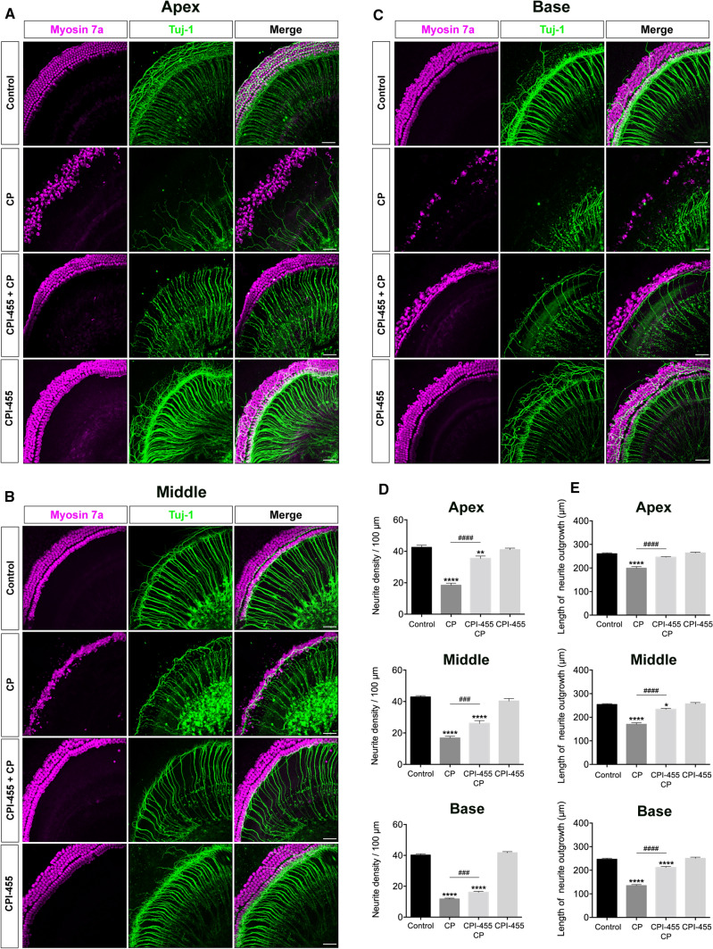
Fig. 4.
CPI-455 reduced CP-mediated SGNs loss in ex vivo cochlear explants. A–C Representative confocal photomicrographs show the spiral ganglion neurons from the apex, middle, and base turns of the cochlea treated with medium alone (control), CP-only (CP), CPI-455 + CP and CPI-455 only for 24 h and stained with Myosin 7a (magenta) and Tuj-1 (green). Scale bar 50 μm. D, E Quantification analysis of neurite densities and the length of neurite outgrowth from the apex, middle, and base turn of cochlear explants for all treatment conditions. The data are presented as the mean ± SEM. *P < 0.05, **P < 0.01, ****P < 0.0001 compared with the control; ###P < 0.001, ####P < 0.0001 compared with CP only. The experiments were performed in triplicate. n = 10 cochleae for control group; n = 10 cochleae for CP group; n = 10 cochleae for CPI-455 + CP group; n = 10 cochleae for CPI-455 group

