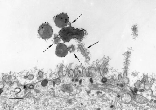
FIG. 5.
TEM demonstrates microvillus engagement (solid arrow) of NTHI 2019 at the surface of a primary airway epithelial cell in an air interface culture after 4 h of infection. Portions of multiple microvilli, surrounding the bacteria (dashed arrows), can be seen in cross section. This specimen was labeled prior to Epon embedment with a rabbit polyclonal antiserum to NTHI 2019, followed by a secondary antiserum conjugated to ultrasmall gold beads. The label was enhanced with silver after embedment and sectioning. The scale bar represents 500 nm.

