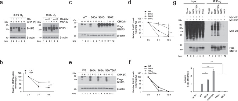
Fig. 3. Phosphorylation of BNIP3 at S60/T66 is essential to improve its stability.
a PC12 cells were exposed to 0.3% O2 supplemented with 200 nM okadaic acid (OA) for 12 h and then treated with 20 μg ml−1 cycloheximide (CHX) for the indicated times (left panel) or with 100, 200 nM OA plus 10 μM MG132 for 6 h (right panel). Cell lysates were detected via western blotting using the indicated antibodies. b Quantification of degradation rate of BNIP3 in CHX chase experiments shown in (a). c–f HeLa cells were transfected with plasmids encoding WT or the indicated BNIP3 mutants for 48 h and then treated with 20 μg ml−1 CHX for 0 h, 6 h, or 12 h. BNIP3 expression was detected via western blotting. d and f are quantification of degradation rate of BNIP3 shown in (c) and (e), respectively. g HeLa cells were transfected with Myc-Ub and empty vector, WT or the indicated BNIP3 mutant for 48 h and treated with 10 μM MG132 for 12 h. Cell lysates were boiled and immunoprecipitated with an anti-Flag antibody. The immune complexes were then analyzed via western blotting. n = 3. The data are expressed as means ± SEM. *P < 0.05, **P < 0.01, ***P < 0.001 versus the indicated group.

