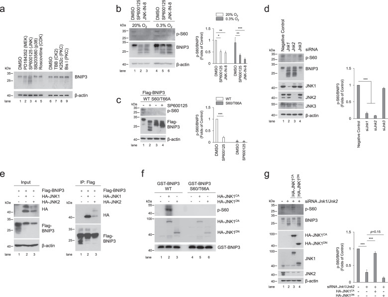
Fig. 5. JNK1/2 is the kinase responsible for BNIP3 phosphorylation.
a PC12 cells were treated with various kinase inhibitors, including 10 μM PD184352 (MEK inhibitor), 10 μM SP600125 (JNK inhibitor), 10 μM SB203580 (p38 inhibitor), 10 μM Roscovitine (CDK inhibitor), 100 μM TBB (CK2 inhibitor), 10 μM K252c and 1 μM Bis I (PKC inhibitor). Then, cell lysates were subjected to western blotting with the indicated antibodies. DMSO, dimethylsulfoxide. b The phosphorylation of BNIP3 was detected via western blotting after PC12 cells were treated with JNK inhibitor SP600125 (10 μM) or JNK-IN-8 (10 μM) and exposed to 20% O2 or 0.3% O2 for 6 h. n = 3. c PC12 cells stably expressing WT or S60/T66A mutant Flag-BNIP3 were treated with DMSO or SP6000125 and followed by detection of phosphorylation of BNIP3. n = 3. d After Jnk was knocked down with the indicated siRNA in PC12 cells, the levels of JNK protein and BNIP3 phosphorylation were measured by western blotting. n = 3. e HeLa cells were transfected with Flag-BNIP3 and HA-JNK1 or HA-JNK2 for 48 h. Cell lysates were then immunoprecipitated with an anti-Flag antibody and detected by western blotting with an anti-HA or anti-Flag antibody. f Immunoprecipitated constitutively active (CA) HA-JNK1 or dominant negative (DN) HA-JNK1 was incubated with WT or S60/T66A GST-BNIP3 protein at 30 °C for 30 min, and then the reaction products were subjected to western blotting with the indicated antibodies. g Jnk1 and Jnk2 knockdown PC12 cells were transfected with HA-JNK1CA or HA-JNK1DN mutants for 48 h, cell lysates were then analyzed via western blotting with the indicated antibodies. n = 3. The data are expressed as means ± SEM. *P < 0.05, **P < 0.01, ***P < 0.001 versus the indicated group.

