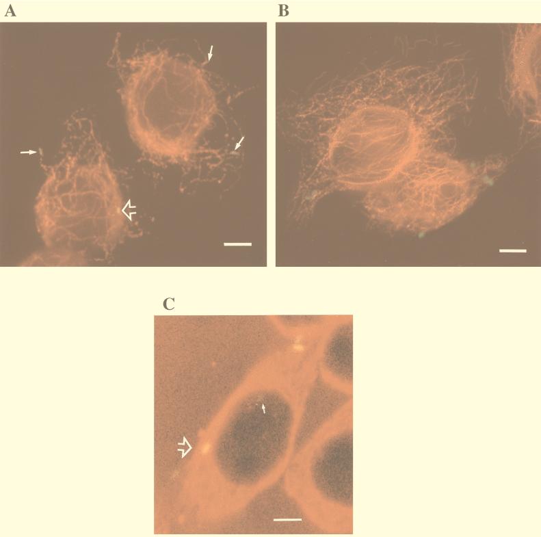
FIG. 4.
Representative microscopic images of immunofluorescently labeled C. jejuni-infected INT407 cells showing specific colocalization of C. jejuni with MT’s. Host cells infected with C. jejuni 81-176 or control S. typhi were prepared and immunolabeled as described in Materials and Methods, using Texas red-X to label MTs and FITC or Oregon Green 514 to label the bacteria. (A) Combined fluorescence image of host cells infected with C. jejuni for 1 h, showing bacteria typically aligned with MTs (arrows). Tight colocalization of C. jejuni with MTs resulted in a yellow fusion color which could be seen to various degrees with different bacteria (arrowhead). (B) Combined fluorescence image of host cells infected with S. typhi for 1 h, showing no apparent specific association of these bacteria with MTs and no color fusion structures. (C) One confocal microscopic laser plane section of 13 total sections of this host cell, showing a faint green non-MT-associated campylobacter (arrow) and one bacterium tightly colocalized with MTs which appears as a yellow fusion color (large arrowhead). Bar markers represent 10 μm.

