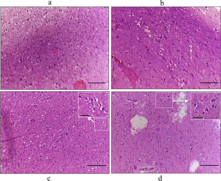
Fig. 1.
Histopathology of FCD types. histopathology of FCD type I and type II: Image was taken using the Olympus microscope. Hematoxylin and eosin staining showed abnormal cortical architecture in FCDI: (a) dyslamination and disorganization in the cortex, b columnar disorganization in the cortex, c FCD IIa showing dysmorphic neurons, and d FCD IIb dysmorphic neurons and balloon cells having large cytoplasm with single or multi nuclei. The scale bar for Fig. 1 a, b, c, and d is 100 µm, and for the high magnification inserts (Fig. 1c, d in white box) 30 µm

