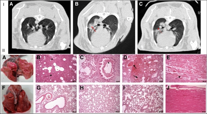
Fig. 2.
I CT scanned images of lung tissue of LPS-induced ARDS in the rabbit. (A) Transverse section of the lung. (B) Inflamed lungs were observed in the control group. (C) Decreased inflammation in the lungs after stem cell treatment. II Necropsy and histopathological findings in the LPS-induced ARDS in the rabbit. (A–E) Control group. (A) Lung tissue is showing hyperemia, hemorrhage, and edema. (B) Interstitial edema and pneumonia (arrow). (B, C) Inflammatory cell infiltration (arrowhead). (D) Hyperemia and severe hemorrhage in alveoli and parenchyma (arrows). (E) Myofibrils necrosis of heart (arrow). (F–K) Treatment group. (F) Lung tissue showing hyperemia and edema (lesser than in the control group). (G–I) Histopathological staining depicts lesser alveoli and parenchymal damage. (K) Heart without injured lesions. Reproduced from [61] and reprinted from Creative Commons Attribution 4.0 licence (CC BY-4.0)

