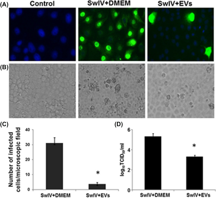
Fig. 9.
SwIV influenza virus replication in lung epithelial cells. A, B Fluorescence and light microscopic images of Lung epithelial cells without influenza virus, Lung epithelial cells incubated with SwIV virus in DMEM media for 1 h, Lung epithelial cells incubated with SwIV virus subjected to 10 µg/ml MSC-EVs treatment for 1 h. C Graph representing the number of viral nucleoproteins expressed cells post 8 h of infection. D Virus titers of SwIV-infected cells and MSC-EV treated cells after 48 h of infection evaluated by titration performed using MDCK cells. Reproduced from [121] and reprinted from Creative Commons Attribution 4.0 licence (CC BY-4.0)

