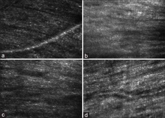
Figure 3.

Images of retinal nerve fiber layer (RNFL) obtained with AOSLO (adaptive optics scanning laser ophthalmoscopy). Images (a and b) belong to the control group and (c and d) belong to patients with early glaucoma. Note that there are no clear differences in the structures of the RNFL between controls and patients with early glaucoma
