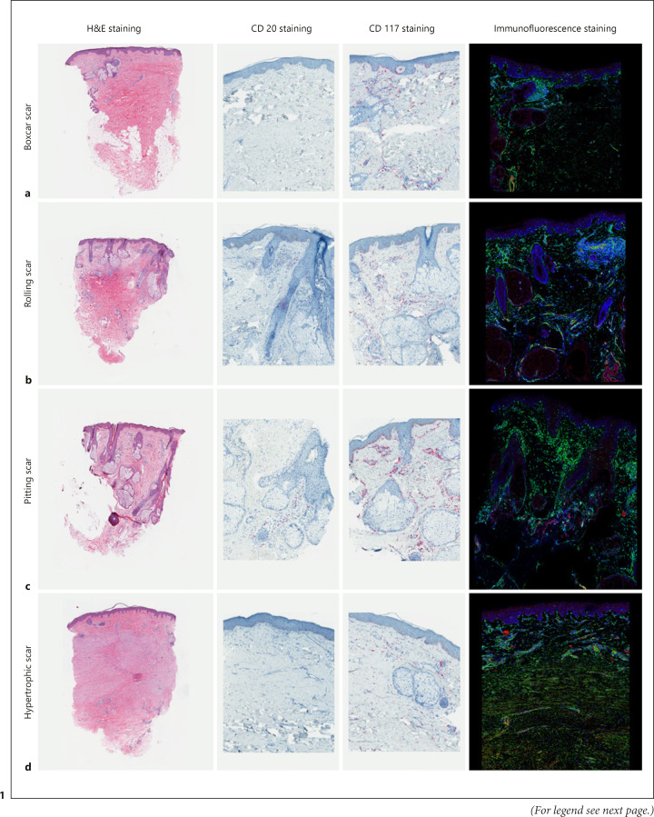
Fig. 1.
Histopathological differences in the examined acne scar specimens. a Boxcar: increased in fibrosis. b Rolling: minimal infiltration of CD117. c Pitting: rich in sebaceous glands. d Hypertrophic: abundant myofibroblasts (yellow cells in immunofluorescence staining). Magnification, ×10 in H&E staining. ×200 in CD20, CD117, and immunofluorescence staining.

