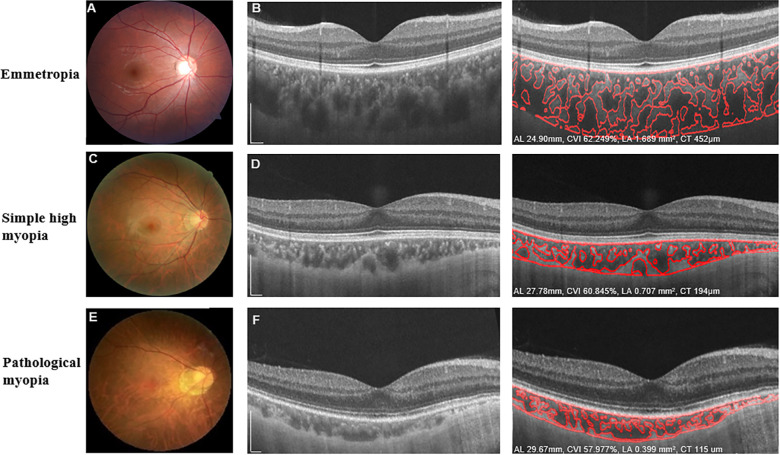
Figure 1.
Characteristic fundus photographs and OCT images of eyes with emmetropia, simple high myopia, and pathological myopia. Fundus photographs (A, C, E) and OCT B-scan images (B, D, F) show the detailed automated segmentation of eyes with emmetropia, simple high myopia, and pathological myopia, respectively. For eyes with emmetropia (A, B), AL was 24.99 mm, the global macular CVI was 62.249%, global macular choroidal LA was 1.839 mm2, and global CT was 452 µm. For eyes with simple high myopia (C, D), AL was 27.78 mm, the global macular CVI was 60.845%, global macular choroidal LA was 0.707 mm2, and global CT was 194 µm. For eyes with pathological myopia (E, F), AL was 29.67 mm, the global macular CVI was 57.977%, global macular choroidal LA was 0.399 mm2, and global CT was 115 µm. Scale bars: 300 µm.

