
FIGURE 1.
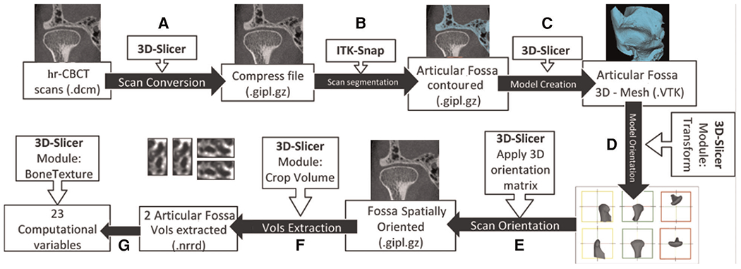
Image processing workflow adapted from bianchi et al. (2021) using 3D Slicer and ITK-SNAP open source software. (A) hr-CBCT files were anonymized and compressed. (B) The condyle and articular fossa were segmented. (C) 3D Slicer was used to convert the segmented articular fossa volume to a 3D surface. (D) Using the “transform” module, a standardized spatial orientation for each 3D TMJ bones model was made. Left TMJ scans were mirrored to the right side. (E) The spatial orientation matrix created in the last step was applied to the TMJ scan. (F) Using the “crop-volume” tool, two regions of the articular fossa (anterolateral, and articular eminence) were selected. (G) Using the “BoneTexture” module, all of the radiomic variables were computed.