
FIGURE 2.
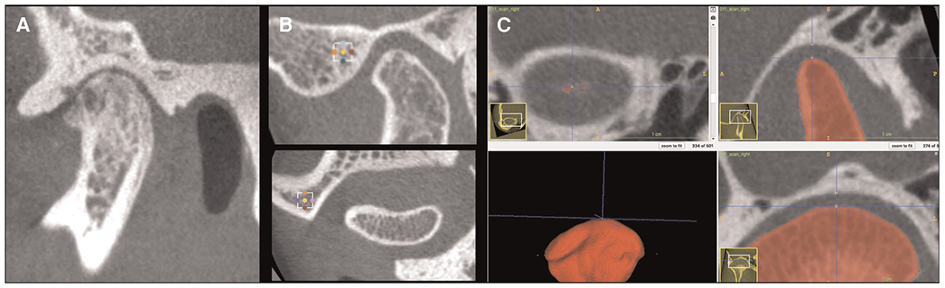
TMJ imaging protocol. (A) Small FOV hr-CBCT scans for TMJ imaging features analysis. Note that marked bone destruction is seen in the condyle and the articular fossa also shows erosion. (B) Volumes of interest (VOIs) in the lateral portion of the articular fossa and in the articular eminence regions. (C) Superior condylar-to-fossa distance as indicated by the blue axis line seen in the coronal and sagittal views.