Fig. 6.
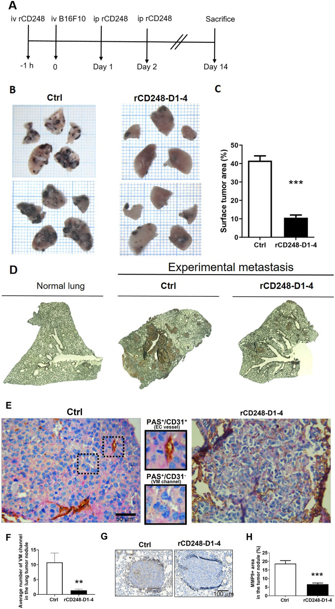
rCD248 inhibits experimental lung metastasis in mice. Experimental lung metastasis assay. A The experimental protocol of mouse model of lung metastasis assay. Twenty µg of rCD248D1-4 protein was given 1 h before and 1 and 2 days after intravenous inoculation of B16F10 cells into each mouse. B Representative pictures of the gross view of mouse lung surface 14 days after tumor inoculation. C Statistical analysis of lung surface occupation of tumor nodules. N = 9 for the control group (Ctrl) and N = 8 for the rCD248D1-4 group. ***, P < 0.001. D Representative CD248 expression patterns in mouse lung isolated from normal mice and mice after experimental metastasis assay. E Representative images of the CD31 (brown color) and PAS (magenta color) double stain in the lung tumor nodules. The PAS+/CD31− area is denoted as vascular mimicry (VM) phenotype. F Statistical analysis of VM channel (PAS+/CD31− stain) in the lung tumor nodule. **P < 0.01. N = 20 nodules for the Ctrl group and N = 21 nodules for the rCD248D1-4 group. G Representative images of MMP9 stain in the lung tumor nodules and H the statistical analysis thereof. ***P < 0.001. N = 117 nodules for the Ctrl group and N = 69 nodules for the rCD248D1-4 group

