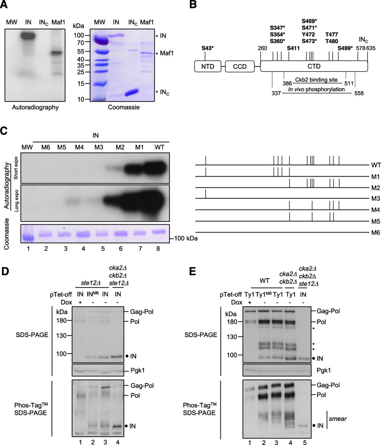
Fig. 2.
CK2 phosphorylates Ty1 integrase in vitro and is involved in its phosphorylation in vivo. A Full-length IN (200 ng), IN578–635 (INC) (500 ng) or Maf1 (200 ng) expressed in E. coli were subjected to in vitro radioactive phosphorylation assays with purified yeast CK2 holoenzyme. Incorporation of γ32P was detected by autoradiography (left panel), the loading of the recombinant proteins was analyzed by Coomassie blue staining (right panel). MW: protein marker. Full length IN, INC and Maf1 are indicated with an asterisk (*). B Mapping of CK2 phosphorylation sites in Ty1 IN. A schematic representation of IN with the N-terminal (NTD), the catalytic core (CCD) and the C-terminal domain (CTD) is depicted [25]. Starting and ending residues for the CTD, the INC sequence and Ckb2 binding site are indicated. All amino acids within a CK2 consensus site listed in Table S4A (Netphos 3.1 analysis) are indicated with an asterisk (*). The 12 amino acids potentially phosphorylated by CK2 in the endogenous IN, based on Phosphogrid database, are shown in bold (Table S4B). C In vitro kinase assays of IN phosphomutants. Left: Autoradiography (incorporation of γ32P) and Coomassie blue staining of WT and mutant IN proteins purified from E. coli (200 ng). MW: protein marker. Right: Mutations preventing phosphorylation of specific residues of IN shown in panel B are indicated by the absence of a vertical line in the representation of IN mutants M1 to M6. D Phosphorylation of ectopically IN expressed alone is not dependent on CK2 in vivo. Total protein extracts from ste12Δ or cka2Δ cbk2Δ ste12Δ cells expressing WT or INM6 (as indicated in panel C) from pTet-off-IN plasmids were separated by SDS-PAGE in the absence (upper panel) or the presence of Phos-Tag™ (lower panel). Pgk1 is a loading control. The presence of IN and Pol intermediates produced from endogenous Ty1 elements was detected by Western blot using rabbit anti-IN polyclonal antibodies. The black circle corresponds to the dephosphorylated form of IN. E Phosphorylation of IN produced from an ectopic Ty1 element depends in part on CK2 in vivo. Western blot analysis as described in panel D of total protein extracts prepared from cells expressing ectopic Ty1 or Ty1M6 mutant from pTet-off plasmids as indicated. Asterisks indicate Pol intermediates from endogenous and ectopic Ty1 elements. IN expressed from pTet-off-IN was used as a control. In Phos-Tag™ SDS-PAGE, the smear above IN indicates the presence of several forms of phosphorylated IN or Pol intermediates. A similar profile was reproduced with cell extracts from 3 independent experiments

