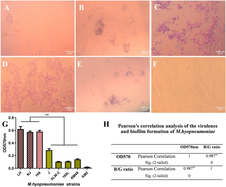
Figure 6.
Biofilm formation ability of M. hyopneumoniae strains. A–F Biofilms formed by strain NJ, which were stained with crystal violet and observed under a microscope after A 1 day, B 2 days, C 3 days, D 4 days, and E 5 days of incubation. F negative control. G, H Comparison of biofilms formed by seven M. hyopneumoniae strains. Biofilm formation ability of strains was determined and identified by crystal violet staining after 4 days of incubation. Bars represent mean ± SD for three independent replicate experiments. KM2 represents control (uninoculated broth).

