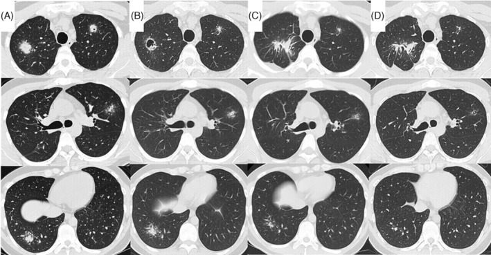
FIGURE 1.

Diagram depicting the timeline of disease progression, CT findings, and management. CT taken at admission shows large nodules with small peripheral nodules, which resemble globular star clusters, in the bilateral upper lobes and right lower lobe (a), formation of cavity in the nodule in the right upper lobe follows (B). One month after starting prednisolone, CT showing the nodules in the left upper and right lower lobes decreased (C). Anti‐human IL‐17A monoclonal antibody is started in January 2021, and prednisolone is terminated in march due to resolution of AS symptoms o. Since then, the nodules in both the left upper and right lower lobes have been gradually decreased (D)
