
FIGURE 2.
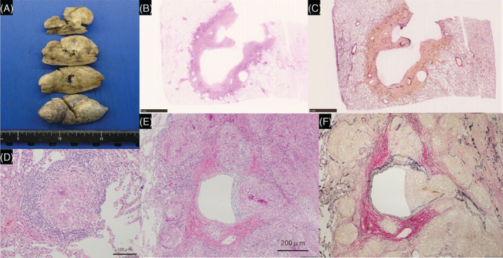
Gross and microscopic histopathology from lung biopsy. Gross view shows a cavity in the resected right upper lobe (A). The image shows granuloma invasion of the vascular walls (B–F), and a large cavity with no airway involvement, suggesting that the cavity is formed as a result of necrosis due to vascular obstruction (E, F). (D: Haematoxylin–eosin, ×200, E: Haematoxylin–eosin, ×100, F: Elastic van Gieson, ×100)