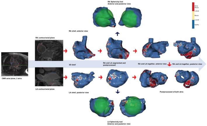
Figure 1. Segmented and postprocessed right and left atria from CMR images.

Steps to obtain the final product for the study. 3D indicates 3‐dimensional; CMR, cardiac magnetic resonance; IIR, image intensity ratio; LA, left atrium; and RA, right atrium.
