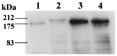
FIG. 3.
Visualization of FnBPs by Western ligand affinity blotting of cell wall-associated protein extracts. Equal amounts of cell wall-associated protein extract were loaded into each lane. Lane 1, S. aureus 8325-4; lane 2, S. schleiferi NCTC 12218; lanes 3 and 4, clinical S. schleiferi isolates (numbered 18 and 3 in Fig. 1, respectively).

