
Figure 5.
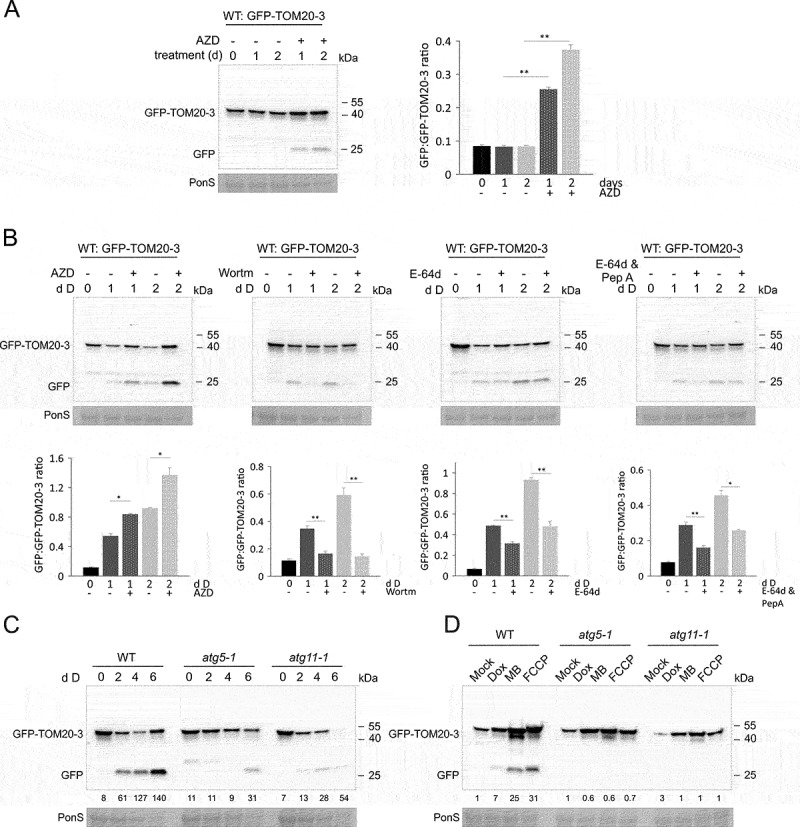
Mitophagy induced during carbon starvation or growth on mitochondria inhibitors is blocked in autophagy mutants, or by chemical autophagy inhibitors. (A) GFP cleavage assay for mitophagy induction by TOR chemical inhibition. 7-d-old GFP-TOM20-3 seedlings grown vertically were transferred to liquid 1/2 MS media, supplemented with 10 μM AZD8055 (AZD) or DMSO and left under Standard light growth conditions for 1 to 2 days. Immunoblot detection of GFP-TOM20-3 and free GFP was performed with anti-GFP antibody. Bar chart on the right represents means (± SE) for free GFP to GFP-TOM20-3 ratios from three independent experiments (Student’s t-test; **p < 0.01). (B) Top panel: Immunoblot detection of GFP cleavage of dark treaded WT seedlings expressing GFP-TOM20-3 using anti-GFP antibody. Total protein extracts were prepared from 7-d-old vertically grown seedlings, incubated for 1–2 additional days in the dark in liquid 1/2 MS media, supplemented with or without 10 μM AZD8055 (AZD), or 5 μM wortmannin (Wortm), or 20 μM E-64d with or without additional supplementation with pepstatin A (PepA). Bottom panel: bar charts illustrating ratios of free GFP to GFP-TOM20-3 from the immunoblots in the top panel. Bars shown are the means (± SE) of three biological replicates (Student’s t-test; *p < 0.05, **p < 0.01). Asterisks denote a significant difference versus seedlings incubated in the dark, without various inhibitors. (C) GFP cleavage assay for carbon starvation (dark-induced senescence) in WT, atg5-1 and atg11-1 seedlings expressing mitochondrial fusion protein GFP-TOM20-3. 10-d-old seedlings grown on 1/2 MS agar medium without sucrose (pH 5.7) were transferred to constant darkness for 2–6 days. Full-length fusion protein and free GFP were detected by immunoblot using anti-GFP antibody. Control Ponceau S stain of RBCL was used to visualize changes in total protein content from lysates upon dark treatment (PonS, bottom panel). (D) GFP cleavage assay of WT, atg5-1 and atg11-1 GFP-TOM20-3 seedlings exposed to mitochondrial chemical inhibitors: Dox, MB and FCCP, for 4 days, using the same experimental system as described in Figure 5D. Presence of the full-length fusion protein and free GFP was confirmed by immunoblot detection with anti-GFP antibody. Ponceau S stained RBCL (PonS) from total protein lysates, was used as a loading control. Relative intensity ratios (% of free GFP:GFP-TOM20-3) on carbon starvation (C) or mitochondrial inhibitors (D) are shown as numeric values.