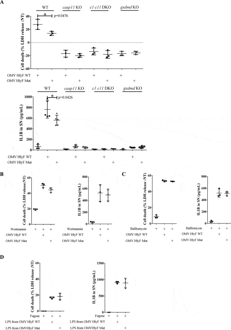
Figure 6.

OMVs HlyF WT induced a stronger activation of the non-canonical inflammasome pathway compared to OMVs HlyF Mut. (A) Release of LDH and IL1B from unprimed WT, casp11−/−, casp1−/−casp11−/− and gsdmd−/− mouse BMDM treated for 24 h with OMV HlyF WT or OMV HlyF Mut 5 µg/mL. The graphs show the mean and the standard deviation of at least 3 independent experiments for each condition. Each dot represents the value obtained in one experiment. *p < 0.05 t test. (B) Release of LDH and IL1B from unprimed WT mouse BMDM treated for 24 h with OMV HlyF WT or OMV HlyF Mut 5 µg/mL with 10 µM wortmaninn. The graphs show the mean and the standard deviation of 3 independent experiments for each condition. Each dot represents the value obtained in one experiment. (C) Release of LDH and IL1B from unprimed WT mouse BMDM treated for 24 h with OMV HlyF WT or OMV HlyF Mut 5 µg/mL with 10 nM bafilomycin. The graphs show the mean and the standard deviation of 3 independent experiments for each condition. Each dot represents the value obtained in one experiment. (D) Release of LDH and IL1B from unprimed WT mouse BMDM transfected with LPS purified from OMVs BL21 HlyF Mut or from OMVs BL21 HlyF WT (1 µg/mL using FuGENE HD, Promega) for 24 h. The graphs show the mean and the standard deviation of 3 independent experiments for each condition. Each dot represents the value obtained in one experiment.
