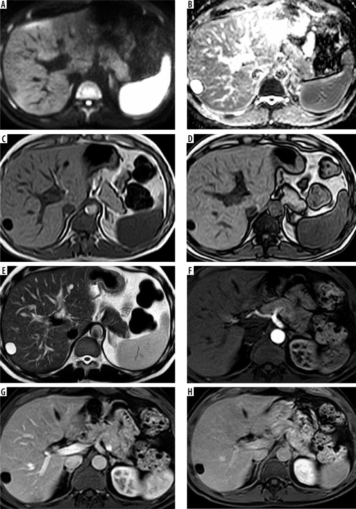
Figure 4.
A 40-year-old male patient with chronic hepatitis C virus (HCV) infection and elevated liver enzymes came for screening. Full standard protocol: A, B) DWI and ADC showed segment VII lesion with no diffusion restriction. C, D) Axial T1WI in phase and out of phase showed a lesion in hepatic subsegment VII eliciting a low signal. E) Axial T2WI: the lesion elicited a high signal. Dynamic contrast study (F, G, H): arterial phase (F), portovenous phase (G), and delayed phase (H) showed no enhancement of the lesion in any of them. The lesion was categorized as LI-RADS-1. Abbreviated 1: (E) Axial T2WI: the lesion elicited a high signal. Dynamic contrast study (F, G, H): arterial phase (F), portovenous phase (G), and delayed phase (H): showed no enhancement of the lesion in all of them. The lesion was categorized as LI-RADS-1. Abbreviated 2: (A, B) DWI and ADC: showed segment VII lesion with no diffusion restriction. Dynamic contrast study (F, G, H): arterial phase (F), portovenous phase (G), and delayed phase (H): showed no enhancement of the lesion in any of them. The lesion was categorized as LI-RADS-1. Abbreviated 3: (C, D) Axial T1WI in phase and out of phase: showed a lesion in hepatic subsegment VII eliciting a low signal. Dynamic contrast study (F, G, H): arterial phase (F), portovenous phase (G), and delayed phase (H) showed no enhancement of the lesion in any of them. The lesion was categorized as LI-RADS-3

