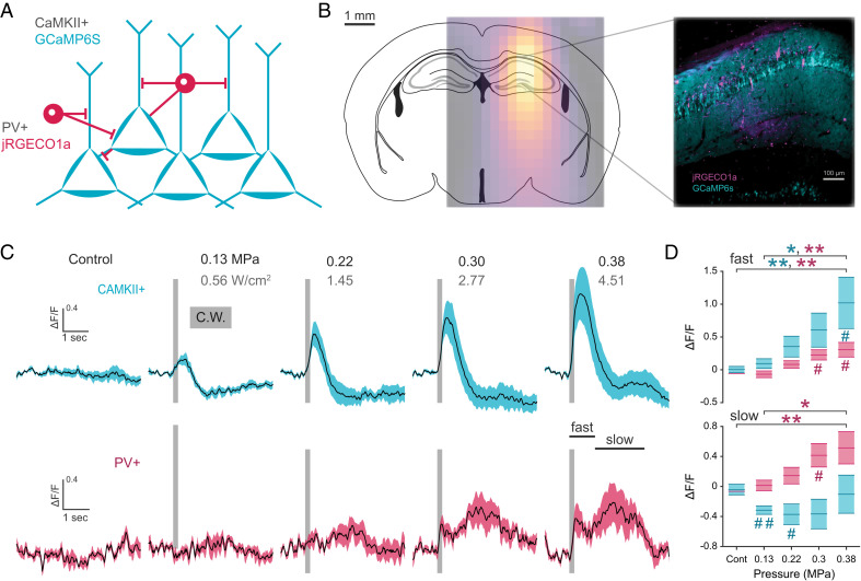
Fig. 2.
Dual cell type labeling reveals temporally and directionally distinct response profiles within the FUS target network. (A) Genetically encoded calcium sensor labeling strategy for CaMKII+ and PV+ neural subtypes of the hippocampus. (B) Overlay of target hippocampal region with ultrasound intensity field and composite image of jRGECO1a and GCaMP6s within the hippocampus. (C, D) Mean cell type change in fluorescence (line) in response to continuous ultrasound pulse (gray bar, 200 ms) of increasing ultrasound pressure (n = 6 animals, shaded area: S.E.M.). Average fast and slow response reveals temporally variant responses between cell types of the CA1 network (one-way ANOVA, Tukey’s multiple comparison test; PV-slow, F4,5 = 6.85; PV-fast, F4,5 = 7.71; CaMKII-slow, F4,5 = 1.49; CaMKII-fast, F4,5 = 5.30; *P < 0.05, **P < 0.01, one-sample t test; #P < 0.05, ##P < 0.01).

