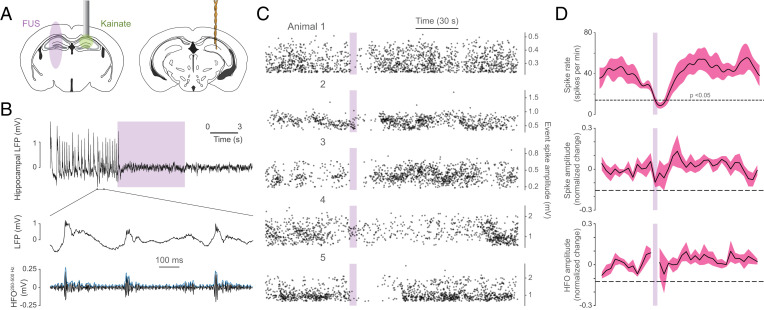
Fig. 5.
900 Hz stimulation of the contralateral hippocampus attenuates epileptiform activity. (A) Illustration of kainate delivery, bipolar electrode, and PhoCUS ultrasound FWHM field strategy. (B) Sample hippocampal local field potential (LFP) shows spontaneous epileptiform spikes accompanied by increases in HFO amplitude. Purple bar indicates ultrasound stimulation. (C) Ten-trial overlay of epileptiform spikes for each animal relative to ultrasound stimulation. (D) Mean spike rate, spike amplitude, and HFO amplitude surrounding stimulation onset (n = 5 animals, dashed line indicates the average P = 0.05 level; two-tailed).

