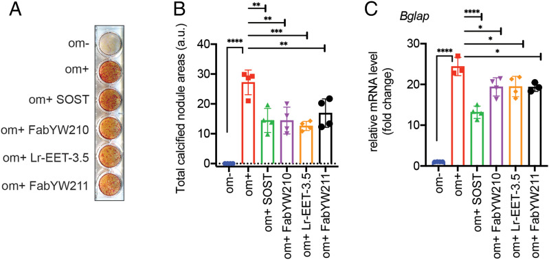
Fig. 5.
Inhibition of osteoblast differentiation. (A) MC3T3-E1 cells were treated with LRP6-binding reagents (1 µg SOST, 1 µM FabYW210 and FabYW211, 5 µM Lr-EET-3.5) for 21 d, and mineralized nodule formation was assessed by alizarin red S staining. FabYW210 binds to the E1-E2 domain of LRP6. The differentiation medium (om+) includes 50 µg/mL ascorbate-2-phosphate and 10 mM β-glycerophosphate (see Materials and Methods). Om- refers to the control medium without the added differentiation factors. (B) Quantification of calcified nodule area. The total calcified nodule area above a threshold defined by the background from om- wells was measured. a.u.: arbitrary unit. (C) Effect on the mRNA expression level of bone marker, osteocalcin (Bglap). The cells were treated for 1 wk before qRT-PCR. Bars represent the means of four independent assays. Error bars represent SDs (*P < 0.05, **P < 0.01, ***P < 0.001, ****P < 0.0001, one-way ANOVA).

