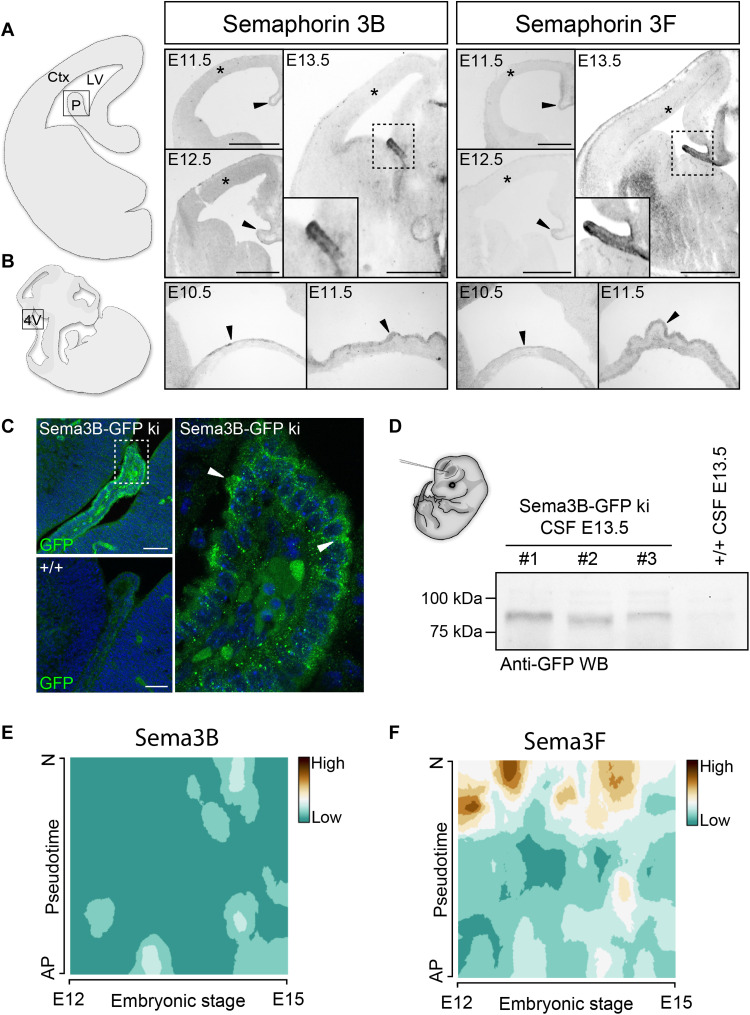
Fig. 1. Class 3 Semas are expressed by the developing CP and secreted into the CSF.
(A and B) In situ hybridizations on coronal sections through the telencephalon (A) and sagittal sections of the hindbrain (B) show the mRNA expression of Sema3B and Sema3F at indicated ages. Arrowheads point to the developing CP. Asterisks indicate the cortical ventricular zone, which is composed of apical progenitors. (C) Fluorescence micrographs showing GFP labeling of the nascent CP in Sema3B-GFP ki/ki and WT embryos at E13.5. Arrowheads point to the GFP accumulation at the apical border of the CP. (D) GFP immunoblotting on CSF from Sema3B-GFP ki/ki embryos shows the presence of soluble Sema3B-GFP molecules in the CSF. (E and F) Mapping of Sema3B (E) and Sema3F (F) mRNA levels obtained from single-cell transcriptome sequencing of embryonic cortical cells between E12 and E15. The “pseudotime” scale indicates the differentiation state of individual cortical cells from an apical progenitor (AP) to a postmitotic neuron (N) deduced from transcriptome analysis. Blue indicates that neither Sema3B (E) nor Sema3F (F) is expressed by cortical progenitor cells. Scale bars, 500 μm (A) and 100 μm (C). Ctx, cortex; LV, lateral ventricle; P, choroid plexus; 4V, fourth ventricle; WB, Western blot.

