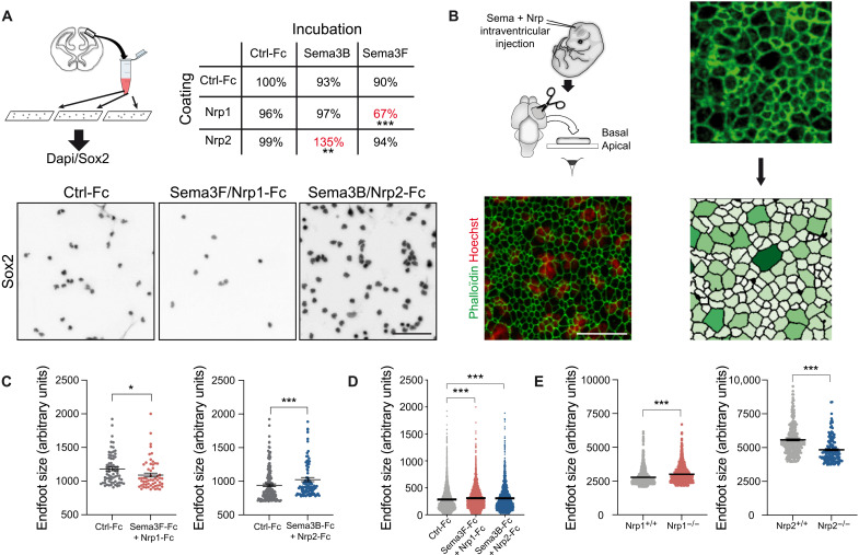
Fig. 4. Dual effects of Sema/Nrp complexes on cortical progenitor adhesion and apical endfeet size in the developing cerebral cortex.
(A) Adhesion assays on coverslips coated with recombinant Nrp-Fc and Sema-Fc molecules in different combinations showed that Sema3F-Fc/Nrp1-Fc inhibited the attachment of Sox2-positive nuclei to the substrate, whereas Sema3B-Fc/Nrp2-Fc promoted cellular attachment. Paired t test, **P < 0.01 and ***P < 0.001. (B) Injections of recombinant Sema3-Fc, Nrp-Fc, or control-Fc were performed in the lateral ventricles of E12.5 mouse embryos. Cortices were labeled with phalloidin–fluorescein isothiocyanate (FITC) and Hoechst. En face confocal images at the levels of apical endfeet and nuclei of apical progenitors were superimposed. The picture illustrates that mitotic cells have the biggest feet (32). The outline of phalloidin-labeled endfeet was detected automatically and the size was measured. (C) Quantitative analysis of the apical endfeet area. Phalloidin-labeled apical endfeet were assorted according to their size, and the largest apical endfeet were plotted. Exposure to Sema3B/Nrp2-Fc results in an augmentation of the 2% largest apical endfeet in comparison to control-Fc. In contrast, Sema3-Fc/Nrp1-Fc conditions decrease the size of the 5% largest apical endfeet. KS test, *P < 0.05 and ***P < 0.001. (D) Totality of analyzed endfeet after intraventricular injection of control-Fc, Sema3-Fc/Nrp1-Fc, or Sema3B/Nrp2-Fc. Means ± SEM; each dot represents one RGC (n = 2 embryos); KS test, ***P < 0.001. (E) Analysis of endfeet size in Nrp1 and Nrp2 mutant mice. Apical endfeet were labeled with phalloidin-FITC and assorted according to their size, and the 5% largest apical endfeet was plotted. Means ± SEM; each dot represents one RGC (Nrp1, n = 4 embryos per condition; Nrp2+/+, n = 4 embryos; Nrp2−/−, n = 2 embryos); KS test, ***P < 0.001. Scale bars, 100 μm (A) and 5 μm (B).

