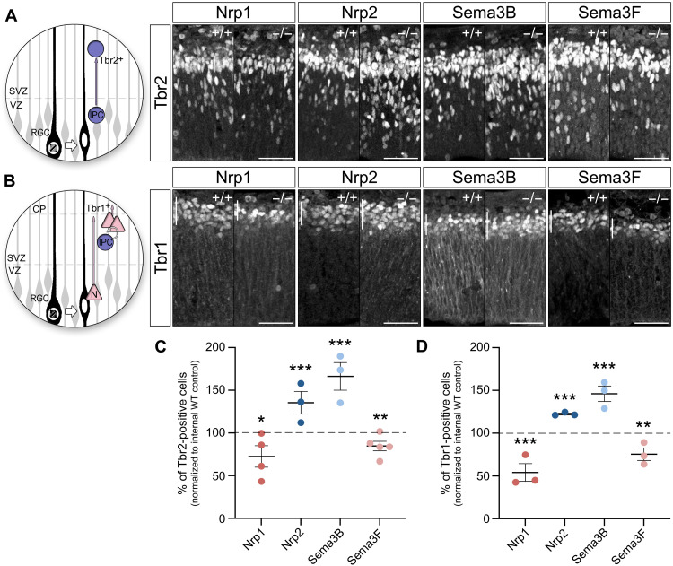
Fig. 6. Sema/Nrp complexes regulate the generation of intermediate precursors and postmitotic cortical neurons.
(A) Tbr2-positive intermediate progenitors delaminate from the apical surface and translocate their soma into the SVZ. Microphotographs illustrate Tbr2-positive nuclei in WT, Nrp1Sema/Sema, Nrp2−/−, Sema3B−/−, and Sema3F−/− embryonic cortices. (B) During early neurogenesis, Tbr1-positive postmitotic neurons are generated directly by radial glia cells or indirectly by intermediate progenitor cells. Microphotographs illustrate Tbr1-positive nuclei in Nrp1Sema/Sema, Nrp2−/−, Sema3B−/−, and Sema3F−/− embryonic cortices. (C) The plot shows that Sema3F−/− and Nrp1Sema/Sema cortices have less Tbr2-positive cells in comparison to the WT littermates at E12.5. In contrast, Sema3B and Nrp2 ko embryos show significant augmented numbers of intermediate progenitor cells. (D) The plot shows that Sema3F−/− and Nrp1Sema/Sema mice exhibit reduced numbers of Tbr1-positive neurons in the cortical plate at E12.5. In contrast, Tbr1-reactive cells are increased in Sema3B and Nrp2 ko mice in comparison to WT littermates. Scale bars, 50 μm. CP, cortical plate; VZ, ventricular zone; SVZ, subventricular zone. Means ± SEM; each dot represents one embryo; paired t test, *P < 0.05, **P < 0.01, and ***P < 0.001.

