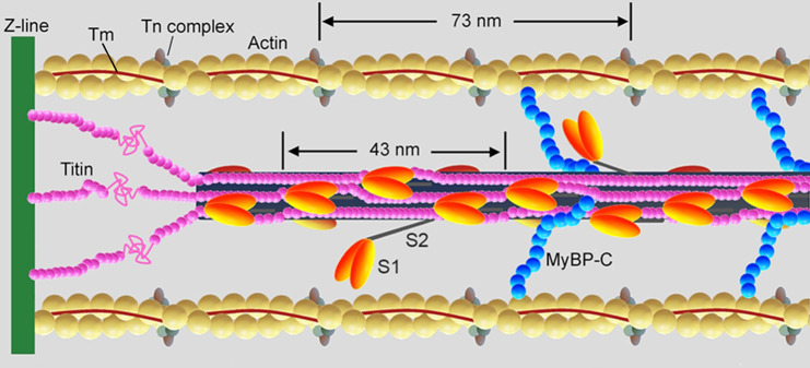
Fig. 1. Schematic representation of the half-sarcomere protein assembly.
Shown are actin (yellow), tropomyosin (Tm, brown) and troponin complex (Tn, light and dark grey and violet) on the thin filament. On the thick filament (black) most of the S1 globular head domains of myosin (orange) lie tilted back (OFF state) and a few heads (two in the scheme) move away with tilting of their S2 tail domain (ON state); the MyBP-C (blue) lies on the thick filament with the C-terminus and extends to thin filament with the N-terminus. Titin (pink) in the I-band connects the Z line at the end of the sarcomere (green) to the tip of the thick filament and in the A-band runs on the surface of the thick filament up to the M-line at the centre of the sarcomere. Adapted from Fig. 1 in ref. 78 (permission from Springer Nature).

