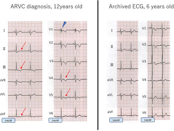
FIGURE 2.

ECG in patient 2. T wave in leads III and V1‐3 was negative at 6 years old (b), but subsequent assessment at 12 years old revealed that those in leads III, aVf, and V1‐4 became negative (a) when marked RV dilatation with suppressed contractility was identified. At this point, LV contractility was preserved and late potentials were positive
