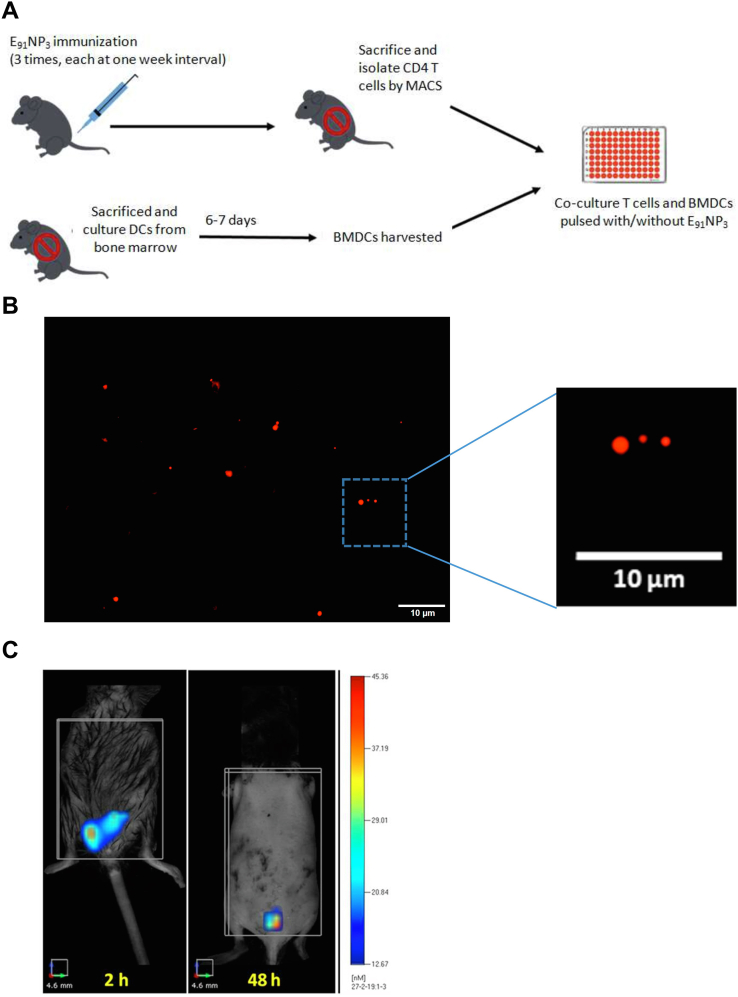
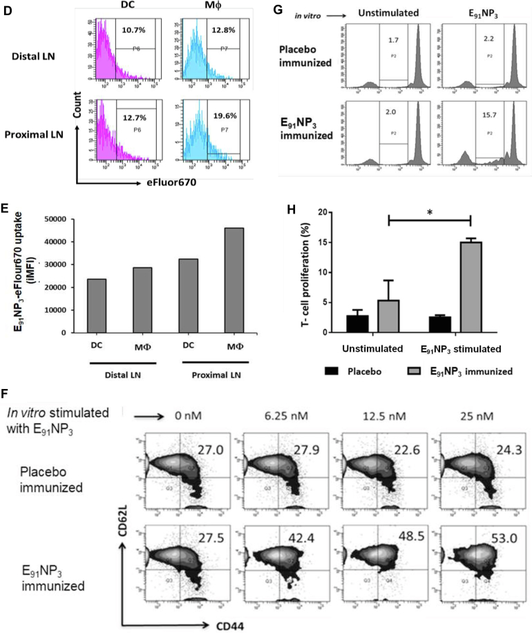
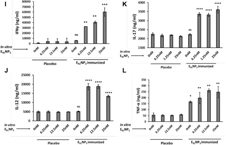
Figure 8.
FMT tomographic live imaging of mice injected with E91NP3-FITC and priming of CD4 T cells by APCs. eFlour670 dye-labeled NPs (E91NP3-eFlour670) were injected s.c. in anesthetized mice. B, fluorescent microscopy images confirm the labeling of NPs with eFluor670. C, FMT images of a C57BL/6 mouse show time-dependent (2 h, 48 h, and 8 days) reduction of fluorescent intensity of E91NP3-eFlour670 from the site of injection. On day 8 postimmunization, inguinal and axillary LNs were processed, and cells isolated were stained and analyzed by flow cytometry. D, histograms represent a percentage. E, bar graph depicts the iMFI changes because of uptake of E91NP3-eFlour670 by DCs and macrophages (Mφ) to axillary (distal) LNs and inguinal proximal LNs from the site of immunization on day 8 post s.c. immunization. A, mice were immunized i.n. with E91NP3, followed by two boosters at intervals of 7 days. Seven days after the second booster, the animals were sacrificed. Splenocytes and LNs were pooled, and CD4 T cells were isolated. CD4 T cells were CFSE-labeled and cocultured with E91NP3 (6.25–25 nM) pulsed DCs. The cells were harvested after 72 h of incubation for flow cytometry analysis, and SNs were collected for the estimation of cytokines. F, contour plots represent CD4 T-cell central memory (CD44hiCD62Lhi) population. G and H, the histogram and bar graph depict the proliferation of CD4 T cells stimulated with or without E91NP3. I–L, the cytokines were estimated in the SNs by ELISA. Bar diagrams represent the level (nanograms per milliliter) of (I) IFN-γ, (J) IL-12, (K) IL-17, and (L) TNF-α. The data are representative of two to three independent experiments with three mice per group. ∗p < 0.05, ∗∗p < 0.01, ∗∗∗p < 0.001, and ∗∗∗∗p < 0.0001. APC, antigen-presenting cell; CFSE, 5(6)-carboxyfluorescein diacetate N-succinimidyl ester; DC, dendritic cell; FMT, fluorescence molecular tomography; IFN-γ, interferon gamma; IL, interleukin; iMFI, integrated mean fluorescence intensity; LN, lymph node; NP, nanoparticle; SN, supernatant; TNF-α, tumor necrosis factor-alpha.


