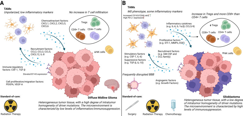
Fig. 3.
Comparison of the immune microenvironment in DMG and GBM. A DMG is found in the midline of the brain and consists of heterogeneous tumor tissue with a high spatial and temporal homogeneity in driver mutations. The microenvironment shows low levels of immune infiltration, inflammation, and immunosuppression. There is no increase in T cell infiltration compared to other pediatric gliomas, while there is a decrease in natural killer (NK) cells compared to healthy children. The TAM population (consisting of microglia and macrophages) is unpolarized, expressing low levels of pro-inflammatory markers. Only selected cytokine/chemokine factors have been confirmed in DMG. The standard-of-care for DMG is radiation therapy. B GBM is found throughout the brain, consists of heterogeneous tumor tissue with low spatial and temporal homogeneity of driver mutations, and often coincides with a disrupted blood–brain barrier (BBB). The microenvironment shows high levels of immunosuppression, characterized by infiltration of Treg T cells. In addition, the ratio of CD8 + to CD4 + T cells and the number of NK cells in the tumor are increased. The TAM population in GBM shows some pro-inflammatory markers and is associated with an M0 phenotype. TAMs show an increase in the CD163/CD68 ratio, with increased PD-L1 expression. Many cytokines and chemokines have been found in GBM samples, of which a selection is shown in the figure. The standard-of-care for GBM is surgery, radiation therapy, and chemotherapy. Created with BioRender.com

