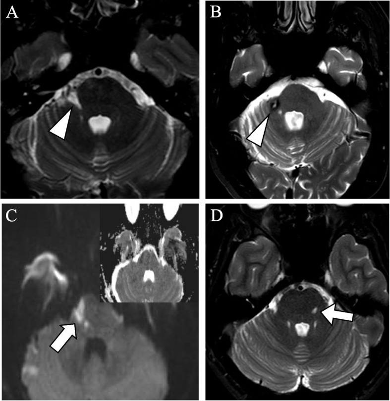
Fig. 3.

Postoperative MRIs of patients with stroke after microvascular decompression. Postoperative MRI of patients after MVD. (a) axial T2 DRIVE weighted sequence shows chronic infarction (arrowhead) in the right cerebellar peduncle of patient no 1 (Supplementary material B). (b) axial T2 DRIVE weighted sequence shows sequelae after hemorrhage (arrowhead) at the right cerebellar peduncle of patient no. 7. c) axial diffusion-weighted sequence shows subacute infarction (arrow) in the right side of the pons of patient no. 3. (d) axial T2 DRIVE weighted sequence shows chronic infarction (arrow) in the left side of the pons in patient no. 4
