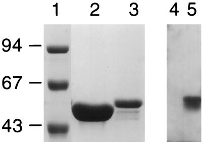
FIG. 1.
Expression of recombinant 9B peptide-1 flagellin protein and its reaction with anti-9B peptide-1 antibodies. Lanes 1 to 3, Coomassie blue staining of purified proteins on sodium dodecyl sulfate–10% polyacrylamide gels. A shift in molecular weight of the recombinant flagellin (lane 3) compared to the native flagellin (lane 2) is seen. Molecular weight markers (in thousands) are shown in lane 1. Lanes 4 and 5, Western blot developed with anti-9B peptide-1–BSA antibodies. No reaction is observed with native flagella (lane 4), while a strong specific reaction is observed with the recombinant 9B-expressing flagella (lane 5).

