
Figure 4.
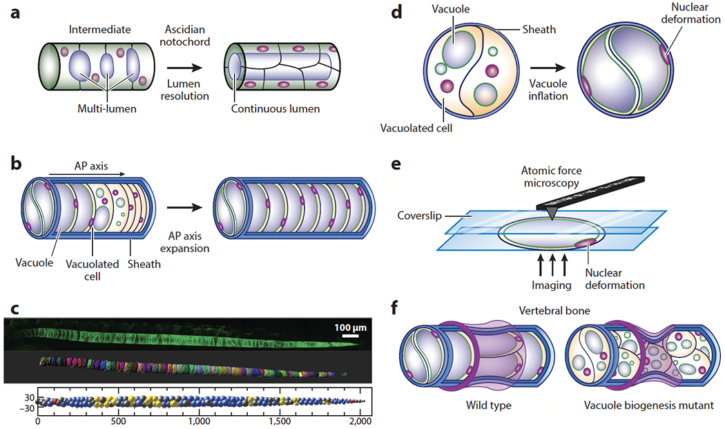
Hydrostatic pressure in the anterior-posterior (AP) axis of chordates. (a) Schematic of continuous lumen formation in the ascidian notochord. Lumen inflation in the notochord provides a hydrostatic scaffold for the free-swimming larva. Note that the multi-lumen intermediate resembles the organization of the vertebrate notochord. (b) Formation and inflation of fluid-filled vacuoles in the vertebrate notochord facilitates AP axis elongation. (c, top) Confocal image, (middle) 3D rendering, and (bottom) 3D arrangement of vacuolated cells in the zebrafish notochord. Vacuolated cells arrange in a stereotypical pattern (see middle panel) dictated by the relative sizes of the cells and the notochord and the aspect ratio of the tube (Norman et al. 2018). Different 3D patterns are color coded. Images in panel c provided by James Norman. (d) Inflation of vacuoles inside vacuolated cells leads to hydrostatic pressure generation inside the semirigid notochord sheath. Nuclear deformation can be used to estimate relative pressure (Bagwell et al 2020). (e) Compression of isolated vacuolated cells using atomic force microscopy and calibration of nuclear deformation may allow the determination of pressure values in vivo via noninvasive imaging. (f, top) Hydrostatic pressure in the notochord resists the compressive force of vertebral bone growth, leading to the formation of symmetrical, hourglass-shaped vertebrae. (Bottom) Loss or fragmentation of vacuoles reduces internal notochord pressure, making the structure more easily deformable and causing vertebral malformations.