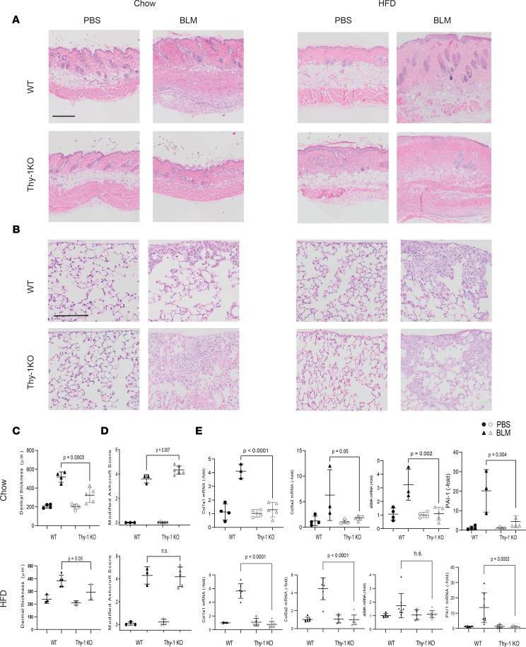
Figure 3. Thy-1–KO mice have attenuated bleomycin-induced skin fibrosis.
(A) Representative images of H&E-stained skin after bleomycin or PBS injections in chow- and HFD-fed mice. Scale bar: 200 μm. (B) Representative H&E-stained lungs after bleomycin or PBS injections in chow and HFD-fed mice. Scale bar: 100 μm. (C) Dermal thickness as determined by 5 high-power fields per mouse (n = 3–5 mice/group). ANOVA with Tukey post hoc test. (D) Fibrosis scores (modified Ashcroft score). Results are mean ± SD from 10 high-power fields per mouse (n = 3–5 mice/group chow diet, n = 5–10 mice/group HFD). Mann-Whitney U test. (E) Expression of Col1a1, PAI-1, ASMA, and Col5a2 assessed by qPCR. Results were normalized to YWHAZ (n = 3–5 mice/group chow diet, n = 5–10 mice/group HFD). ANOVA with Tukey post hoc test.

