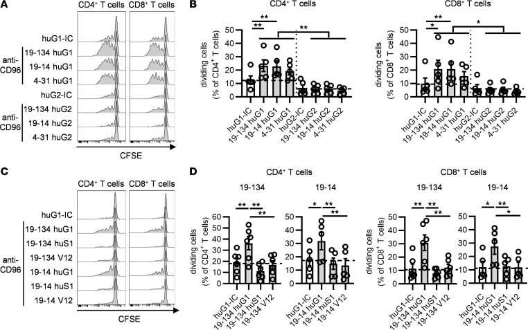
Figure 3. The activity of CD96 mAbs requires FcγR cross-linking.
CFSE-labeled PBMCs from HDs were stimulated for 4 days with soluble OKT3 and soluble anti-CD96 mAb variants as indicated, and the proportion of proliferating cells was determined by flow cytometry. (A and B) The effect of human IgG1 (huG1) and human IgG2 (huG2) anti-CD96 mAbs on CD4+ and CD8+ T cell proliferation was compared. (C and D) The effect of huG1, Fc-silent N297S human IgG1 (huS1), and V12 human IgG1 anti-CD96 mAbs on CD4+ and CD8+ T cell proliferation was compared. (A and C) Representative examples of CFSE dilution. (B and D) Data show the mean ± SEM of the frequency of dividing cells, with each symbol representing the mean of triplicate wells for an individual donor and the dotted lines indicating the percentage of dividing cells after stimulation with the isotype controls. Data are combined from (B) n = 4 independent experiments and (D) from n = 4 and n = 3 independent experiments for clones 19-134 and 19-14, respectively. *P ≤ 0.05, **P ≤ 0.01. One-way ANOVA.

