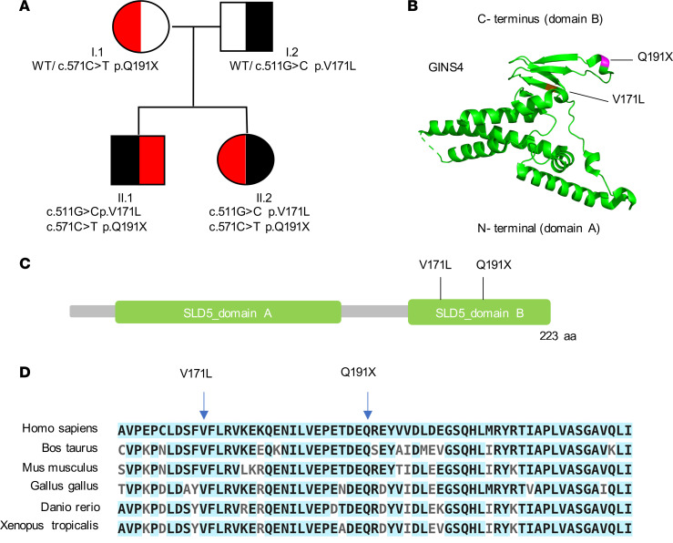
Figure 2. Identification of compound heterozygous variants in the GINS4 gene by whole-exome sequencing.
(A) Pedigree of the family denoting variants of GINS4. (B) GINS4 protein 3D structure prediction showing α-helices at N-terminus and B-strands at C-terminus (variants labeled in violet). (C) Schematic representation of GINS4 (SLD5) protein and variants mapping at the C-terminal domain. (D) Multiple protein sequence analysis using ClustalW shows evolutionary conservation of the C-terminal domain of GINS4. Conserved amino acids are highlighted in light blue. The position of both variants is indicated.

