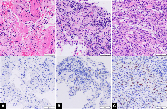
Fig. 3.

Kaposi Sarcoma. A Fine needle biopsy remnant tissue and blood clot showed minute unremarkable lung tissue with no diagnostic tissue (Top panel: Hematoxylin & Eosin stain, 200 × magnification; Bottom panel: HHV-8 immunohistochemistry, 200 × magnification); aspirate smears were acellular (not pictured) B Forceps biopsy showed one fragment of rare spindle cells with crush artifact and focal, weak HHV-8 expression (Top panel: Hematoxylin & Eosin stain, 200 × magnification; Bottom panel: HHV-8 immunohistochemistry, 200 × magnification) C Cryobiopsy showed Kaposi sarcoma with strong and diffuse HHV-8 expression (Top panel: Hematoxylin & Eosin stain, 200 × magnification; Bottom panel: HHV-8 immunohistochemistry, 200 × magnification)
