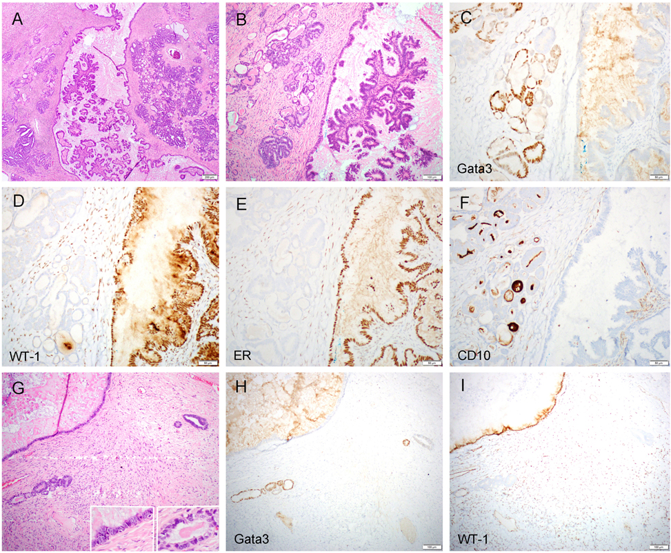
Figure 4.
Serous borderline tumor/atypical proliferative serous tumor (SBT/APST) with mesonephric-like differentiation/hyperplasia (case 2). The left ovarian tumor demonstrated conventional type SBT/APST and a few areas (each area measured less than 5 mm) of mesonephric-like differentiation/hyperplasia (A and B). The former component was negative for Gata3 (C) but diffusely positive for WT-1 (D) and ER (E), while the latter component was focally positive for Gata3 (C) but negative for WT-1 (D) and ER (E). A luminal pattern of CD10 staining was present in the mesonephric-like tubules but not in the SBT/APST (F). In some areas, individual mesonephric-type glands or few glandular clusters were seen in close proximity to the serous-type epithelium (G; left inset: serous epithelium; right inset: mesonephric-like epithelium), displaying distinct immunoprofile for each component (H: Gata3; I: WT-1).

