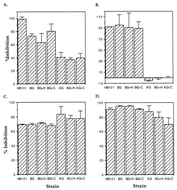
FIG. 3.
Comparison of the ability of cytochalasin D (A), 30-min preincubation with nocodazole (B), colchicine (C), and 2-h preincubation with nocodazole (D) to inhibit the entry of BCYE-grown (BG) and amoeba-grown (AG) L. pneumophila into THP-1 cells. The ability of each of these pharmacological agents to inhibit entry is reported as the percentage of CFU that enter in the presence of the inhibitor compared to a control in the absence of the inhibitor. E. coli HB101 was used as a conventional phagocytosis control. Serum was replaced with RPMI in the no-serum controls (BG and AG). The abbreviation +H indicates that heat-inactivated serum was incubated with the bacteria prior to the assay, and +C indicates that complete human serum was used. % Inhibition = 100 − [100 × (cell-associated CFU in the presence of the inhibitor/cell-associated CFU in the absence of the inhibitor)]. Error bars represent the standard deviations of triplicate wells for each experiment. The results shown are from a single representative experiment of at least three independent experiments.

