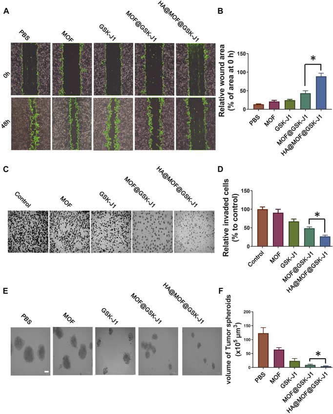
FIGURE 5.
Effects of HA@MOF@GSK-J1 on CR SKOV-3 cell migration, invasion, and self-renewal. (A) Confluent CR SKOV-3 cells were scratched and incubated with MOF, GSK-J1, MOF@GSK-J1, and HA@MOF@GSK-J1, respectively. The area covered by the migrating cells was imaged at 0 and 48 h. (B) Rates of wound closure in wound scratches receiving different treatments. (C) CR SKOV-3 cell invasion tested by Boyden chamber assays. Media containing 0.1% FBS (−) was used as the negative control. (D) Quantification of migrated CR SKOV-3 cells after incubation with MOF, GSK-J1, MOF@GSK-J1, and HA@MOF@GSK-J1, respectively. Tumor suppressor effects of GSK-J1 and HA@MOF@GSK-J1 NPs were observed when CR SKOV-3 cells were cultured as tumor spheroids in 3D assay platforms. (E) Representative images of tumor spheroids after incubation with MOF, GSK-J1, MOF@GSK-J1, and HA@MOF@GSK-J1, respectively, for 3 days. Scale bar: 100 μm. (F) Tumor spheroid volumes after treatment with MOF, GSK-J1, MOF@GSK-J1, and HA@MOF@GSK-J1. Data are presented as means ± SD of three independent experiments. *p < 0.05.

