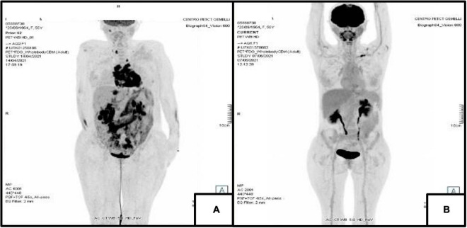
FIGURE 2.
18F-FDG Positron Emission Tomography. (A) PET/CT of April 2021 showing disease with intense metabolic activity in the following areas: SVC, aortic root, pulmonary artery, both atria, IAS, IVS and LV in the thorax and liver, both kidneys, mesenterial adipose tissue, annexes and several lymph nodes in the abdomen. (B) PET/CT of June 2021 showing an almost complete resolution of pathological metabolic activity both in the thoracic and abdominal previous sites of uptake.

