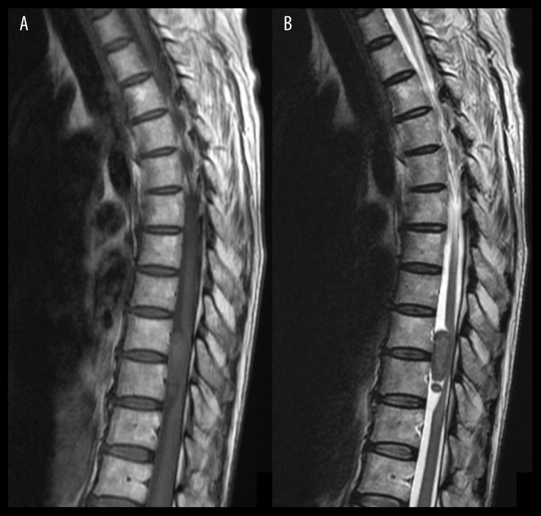
Figure 3.
Thoracic spine magnetic resonance imaging showing an intraspinal mass. Sagittal T1-weighted (A) and T2-weighted (B) images show a mass extending from T9 to T10 with associated significant compression of the spinal cord. No extension to the neuroforamina or associated bony scalloping or bony erosion is seen.

