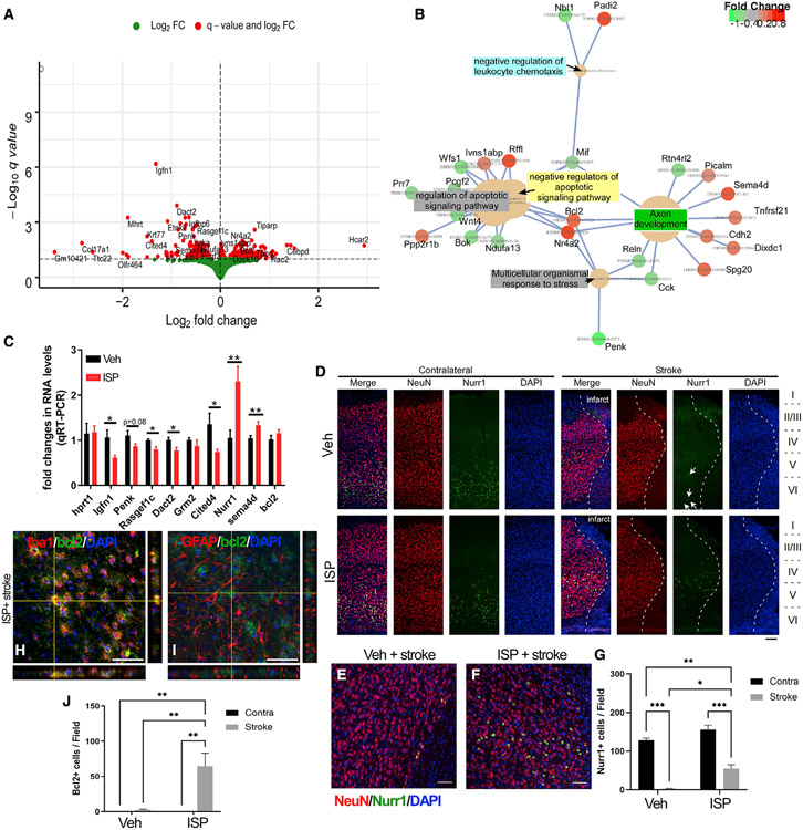
Figure 5. RNA-seq from the peri-infarct cortex of ISP treated versus Veh-treated stroke mice shows differentially regulated genes.
(a) DEGs that can be clustered into GO pathways (B) such as regulators of apoptotic signaling pathways, axon development pathways, and pathways that are involved in responses to stress.
(C) Validation of the top selected gene expression by qRT-PCR (n = 7 for each group).
(D–G) Nurr1 (Nr4a2) expression is decreased in peri-infarct cortex in stroke mice (arrows in D) but partially restored in ISP-treated mice (n = 4 for each group). Nurr1 expression is mainly detected in the NeuN+ neurons in the peri-infarct zone.
(H–J) Bcl2 expression is upregulated in ISP-treated peri-infarct zone enriched in Iba1+ cells not GFAP+ reactive astrocytes (n = 4 for each group). *p < 0.05, **p < 0.01, ***p < 0.001, Student’s t test for (C) and 2-way ANOVA for (J) and (G). Scale bar=50 μm.
See Table S2 for complete list of DEGs.

