Fig. 6.
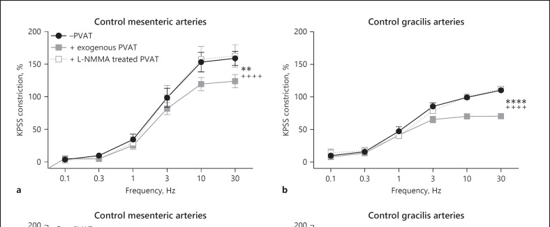
EFS-induced PVAT anticontractile effect is NOS dependent and can be reduced by inhibition of nNOS. Mesenteric (a) and gracilis (b) −PVAT arteries from control mice with a section of exogenous PVAT suspended above were stimulated with EFS. The exogenous PVAT was removed and incubated for 30 min with L-NMMA (100 µM), before returning the PVAT to the organ bath and repeating the stimulus (n = 8 both groups. a −PVAT vs. exogenous PVAT p < 0.01**, exogenous PVAT vs. L-NMMA-treated PVAT p < 0.0001++++, −PVAT vs. L-NMMA-treated PVAT p > 0.05. b −PVAT vs. exogenous PVAT p < 0.0001****, exogenous PVAT vs. L-NMMA-treated PVAT p < 0.0001++++, −PVAT vs. L-NMMA-treated PVAT p > 0.05). Mesenteric (c) and gracilis (d) −PVAT arteries from control mice with a section of exogenous PVAT suspended above were stimulated with EFS. The exogenous PVAT was removed and incubated for 30 min with vinyl-L-NIO (100 nM), before returning the PVAT to the organ bath and repeating the stimulus (n = 8 both groups. c −PVAT vs. exogenous PVAT p < 0.0001, exogenous PVAT vs. vinyl-L-NIO-treated PVAT p < 0.0001****, −PVAT vs. vinyl-L-NIO-treated PVAT p < 0.01**. d −PVAT vs. exogenous PVAT p < 0.0001, exogenous PVAT vs. vinyl-L-NIO-treated PVAT p < 0.0001****, −PVAT vs. vinyl-L-NIO-treated PVAT p < 0.0001****). Data shown are mean ± SEM (−PVAT vs. +PVAT vessels were tested using two-way ANOVA. Before and after drugs within the same vessel type, e.g., +exogenous PVAT before drug vs. +exogenous PVAT after drug were tested using repeated-measures ANOVA. Both followed by Bonferroni post hoc tests).

