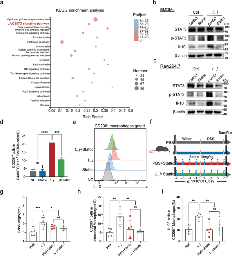
Figure 4.

STAT3 signaling was essential for L. johnsonii activated CD206+macrophagesIL-10.
a, KEGG enrichment analysis of macrophages related gene sets. The degree of color represented p adjust value and the size of node represented the gene number in this item. b-c, The protein expression of STAT3, p-STAT3, Il-10 was tested by western blot in both BMDMs and Raw264.7 cells. d, Percentage of CD206+ macrophages were analyzed by flow cytometry after BMDMs co-cultured with PBS control (NC), Stattic, L. johnsonii (L. j) or Stattic and L. johnsonii (L. j+ Stattic) (MOI = 100:1) for 24 hours. e, Representative histograms of Il-10 expression by CD206+ macrophages were shown after BMDMs co-cultured with PBS control (NC), Stattic, L. johnsonii (L. j) or Stattic and L. johnsonii (L. j+ Stattic) (MOI = 100:1) for 24 hours. f, Schematic diagram showing that the process of STAT3 inactivation model in vivo. g, Colon length was analyzed in four groups. h, Percentage of CD206+ macrophages were analyzed from colon lamina propria in STAT3 inactivation model. i, Percentage of Il-10+ cells were analyzed from CD206+ macrophages in STAT3 inactivation model. Data are presented as mean ± SD, n = 3–5. *, P < .05; **, P < .01; ***, P < .001; ****, P < .0001; ns no significant. ANOVA test (d, g-i).
