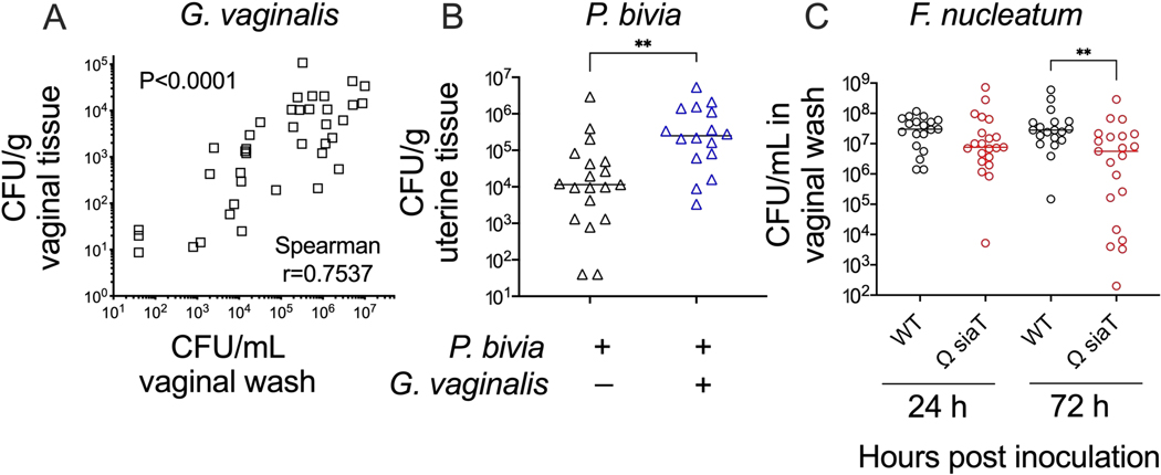
Figure 4: Further analysis of anaerobic bacterial colonization in a murine vaginal inoculation model.
(A) G. vaginalis titers in vaginal washes correlate with G. vaginalis titers recovered from vaginal tissue homogenates at 24 h and 72 h post-inoculation (combination of two independent experiments, n = 10 each. Significance determined by Spearman’s rank correlation test)24. (B) Co-infection with G. vaginalis leads to increased P. bivia titers in uterine horn tissue when compared to P. bivia mono-infected animals at 48 h post-inoculation (combination of two independent experiments, each with 6–10 mice per group. Significance determined by Mann-Whitney U-test, **p = 0.0017)25. (C) F. nucleatum mutant with disrupted sialic acid transporter (Ω SiaT) has decreased titers in vaginal lavages of mono-infected mice as compared to F. nucleatum wild-type at 72 h post-inoculation (combination of two independent experiments, each with 10 mice per group. Significance determined by Mann-Whitney U-test, **p < 0.01)26.

