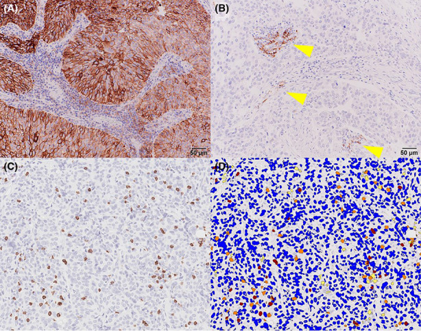
FIGURE 1.

(A) PD‐L1 expression was regarded as positive. (B) PD‐L1 expression was considered to be negative. Only immune cells outside the tumor area expressed PD‐L1 (arrowheads). (C) Pre‐analytical image of CD8 immunohistochemistry. (D) Post‐analytical image shown in Figure 1C. Blue, yellow, orange, and red pixels indicate negative, weak‐positive, positive, and strong‐positive pixels of CD8 immunohistochemistry, respectively
